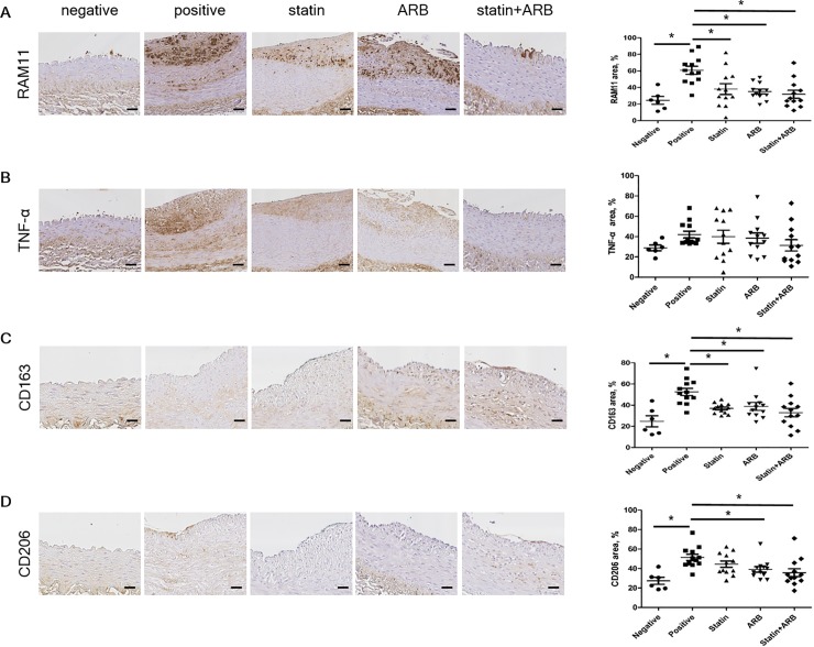
Fig 3. Immunohistochemical staining of macrophage markers.
(A) Tissues immunologically stained with RAM11 and percentage area. (B) Tissues immunologically stained with TNF-α and percentage area. (C) Tissues immunologically stained with CD163 and percentage area. (D) Tissues immunologically stained with CD206 and percentage area. Scale bars = 100 μm. Data are mean±SEM. * p<0.05 vs. positive control group. ARB, angiotensin II receptor blocker; CD163, cluster of differentiation 163; CD206, cluster of differentiation 206; TNF-α, tumor necrosis factor-α.

