Figure 3.
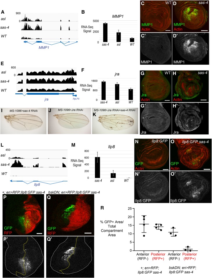
Centrosome loss leads to upregulation of expression of JNK target genes. (A and E) Browser shots of RNA-Seq signals (normalized read depth) of two known JNK target genes, MMP1 (A) and Jra (E), for asl, sas-4, and WT genotypes. Transcription direction indicated by arrowheads. (B and F) Bar plots of average RNA-Seq signals (normalized counts ± SD) of the three biological replicates for MMP1 (B) and Jra (F). (C-C’) MMP1 protein, as visualized by antibody, has minimal expression in WT wing discs. (D-D’) MMP1 protein levels are dramatically increased in acentrosomal sas-4 wing discs. (G-G’) Jra is weakly expressed in control discs, though there is significant expression in the peripodial cells (not shown in this single-slice image). (H-H’) Jra protein is increased in sas-4 discs. (I–K) Representative adult wings from the indicated genotypes. Neither knockdown of sas-4 alone (I) nor jra alone (J) perturbs wing development. However, knockdown of both sas-4 and jra together produces necrotic spots in the adult wing. (L) Browser shot of RNA-Seq signal (normalized read depth) of the ilp8 locus for asl, sas-4, and WT genotypes. (M) Bar plot of average RNA-Seq signal (normalized counts ± SD) for ilp8 in the three genotypes. (N-N’) ilp8 expression, as assessed using a protein trap line expressing GFP-tagged Ilp8 under control of the ilp8 promoter, is low in control discs. (O-O’) ilp8 is upregulated in sas-4 discs. (P) A sas-4 mutant wing disc expressing Ilp8:GFP and en>RFP with no transgene, is a control for the experiment in Q (P’ shows the Ilp8:GFP channel alone). (Q) The upregulation of ilp8 associated with centrosome loss is JNK-dependent because misexpression of BskDN in the posterior portion of sas-4 homozygous mutant wing discs inhibits Ilp8:GFP upregulation. BskDN is driven by en>RFP (red in Q; grayscale in Q’ is the Ilp8:GFP channel alone). White dashed line marks the outer edge of the wing disc. (R) Quantification of Ilp8:GFP positive area standardized to the total area of the anterior (RFP negative) or posterior (RFP positive) area. Note that expression of en>RFP alone does not alter Ilp8:GFP levels induced by centrosome loss, whereas en>RFP driving bskDN noticeably reduces Ilp8:GFP levels. Bar, 50 µm. Images are maximum-intensity projections, except in (G and H) where single slices were used to limit the Jra signal from the peripodial cells. RFP, red fluorescent protein; RNA-Seq, RNA-sequencing; WT, wild-type.

