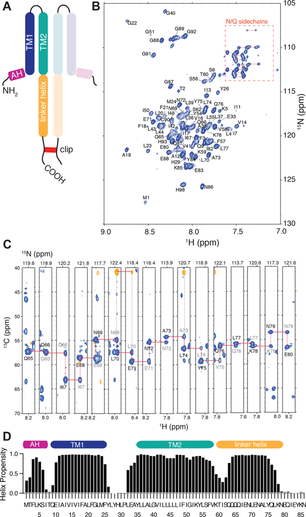
Figure 3.

NMR characterization of the NsaS transmembrane bundle. (a) A schematic of the protein construct used for NMR studies. The construct includes the transmembrane domain and the intracellular linker domain of NsaS. A cysteine tag is attached to the C-terminal to ensure detergentindependent dimerization. (b) Fully assigned 2D 1H−15N TROSY spectrum and (c) 3D strip plot showing sequential backbone assignments of 2H−13C-15N labeled NsaS in 2H C12 betaine micelles at pH 6 (see Methods). Spectra were collected at 900 MHz using a cryo-probe set to 42 C. (d) Chemical shift based secondary structure predictions for NsaS show a helical break at residue 8, an extracellular loop between residues 25–33 and a second helical break around residue 60.
