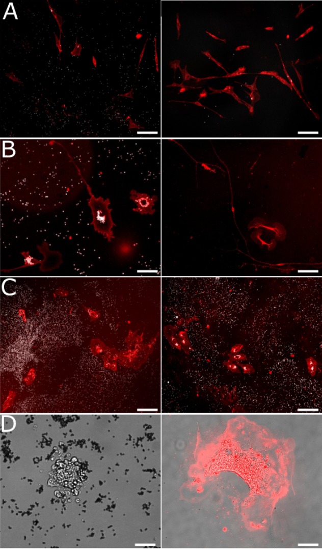
Figure 4. Cells remain viable and proliferate after capture.
Images showing scMOCa-captured cells stained with WGA-Alexa-647. (A) LF-1 fibroblasts 1 (left) and 4 (right) days after sorting. Scale bar 80 µm. (B) Primary DRG neurons 2 (left) and 4 (right) days after sorting. Scale bars: 25 µm (left) and 80 µm (right) (C) HUVECs 3 (left) and 6 (right) days after sorting. Scale bar: 80 µm. (D) Mouse embryonic stem cells 7 days after sorting (left) and 5 days after starting differentiation (15 days after sorting) (right). Prior to differentiation, only a bright-field image is shown to preserve cell viability. After differentiation, we stained cells with WGA-Alexa647, and merged the image with a bright-field photo to increase contrast and better see cellular extensions. Scale bar: 40 µm.

