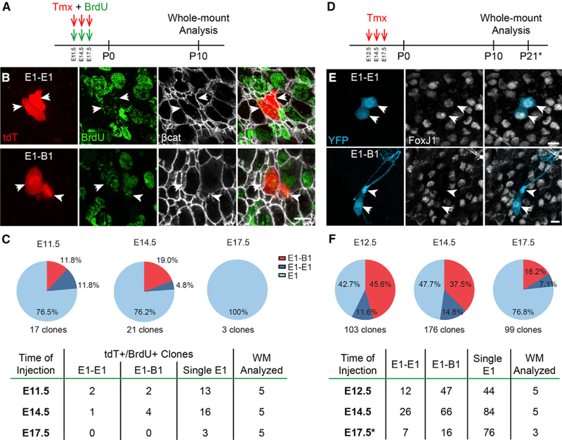
Figure 5. Clonally Related V-SVZ Cells Derived from Nestin+ RG.

(A) Low doses of tamoxifen were given to Nestin::CreER;Ai14 timed-pregnant female mice at E11.5, E14.5, or E17.5. BrdU was injected simultaneously and 6 h later. Whole mounts were analyzed at P10 for double-labeled tdT+/BrdU+ cells.
(B) Confocal images of an isolated tdT+/BrdU+ clone of E1 cells (arrows, top row) and a clone containing a B1 cell (arrowhead, bottom row) and an E1 cell (arrow, bottom row).
(C) Quantification of tdT+/BrdU+ clones observed at different ages of injection, from a total of 5 analyzed whole mounts (WMs) per age.
(D) Sixty-fold higher doses of Tmx were administered to Nestin::CreER;Confetti timed-pregnant female mice at E12.5, E14.5, and E17.5. Whole mounts were analyzed at P10 (E12.5, E14.5) or P21 (E17.5) for single-fluorophore-labeled pairs of cells that included E1 cells (arrows) and/or B1 cells (arrowheads).
(E) Confocal images of isolated single-fluorophore-expressing clones (YFP shown here) of E1 cells (arrows, top row) and of a B1 cell (arrowhead) with an E1 cell (arrow, bottom row).
(F) Quantification of clones observed using the Nestin::CreER;Confetti mice at different ages of injection, from a total of 3–5 WMs analyzed per age.
