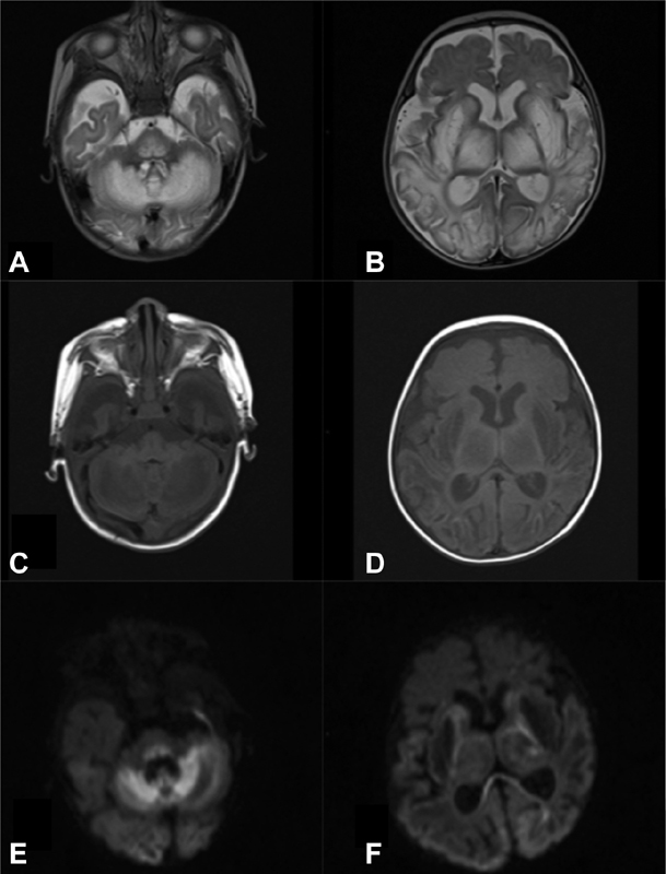
Fig. 1.

Brain MRI (patient 1, at the age of 2 months): Bilateral and symmetrical increased signal intensity in axial T2-weighted images ( A,B) with decreased signal intensity in T1-weighted images ( C, D ) in basal ganglia, thalamus, midbrain, dorsal pons, medulla oblongata, frontotemporal-parieto-occipital cortex, and subcortical white matter and cerebellum. Diffusion weighted imaging: b1000 images ( E, F ) showing extensive areas of restricted diffusion (hyperintensity) in cerebral white and gray matter structures (splenium of corpus callosum, cortex, cerebellum, dorsal pons, and midbrain). Notice the ventricular enlargement and corpus callosum hypoplasia. MRI, magnetic resonance imaging.
