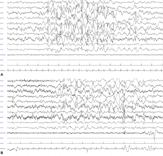
Fig. 4.

( A ) EEG: 30 mm/sec, 14 µV/mm. Burst of generalized polispikes-wave with maximal amplitude over the right central regions, alternating with period of poor low-voltage activity (burst-suppression pattern). ( B ) EEG: 30 mm/sec, 20 µV/mm. Subcontinuouspolispike activity with inter-burst periods of less than 10 seconds, characterized by asynchronous, low-voltage slow activity and multifocal spikes
