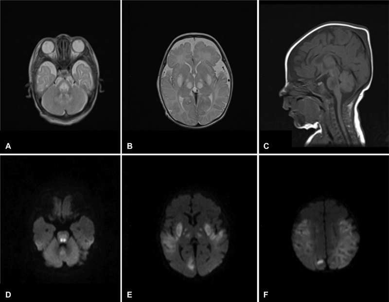
Fig. 5.

Brain MRI (patient 2, at the age of 1 month): Axial T2-weighted images ( A,B ) showing several areas of bilateral symmetric abnormal signal intensity, with diffusion restriction ( D–F ) in the lentiform nuclei, internal capsule, thalami, brainstem, dentate nuclei, and cortical and subcortical regions. Sagittal T1-w images (C) show hypointensity in the affected areas and a thin corpus callosum.
