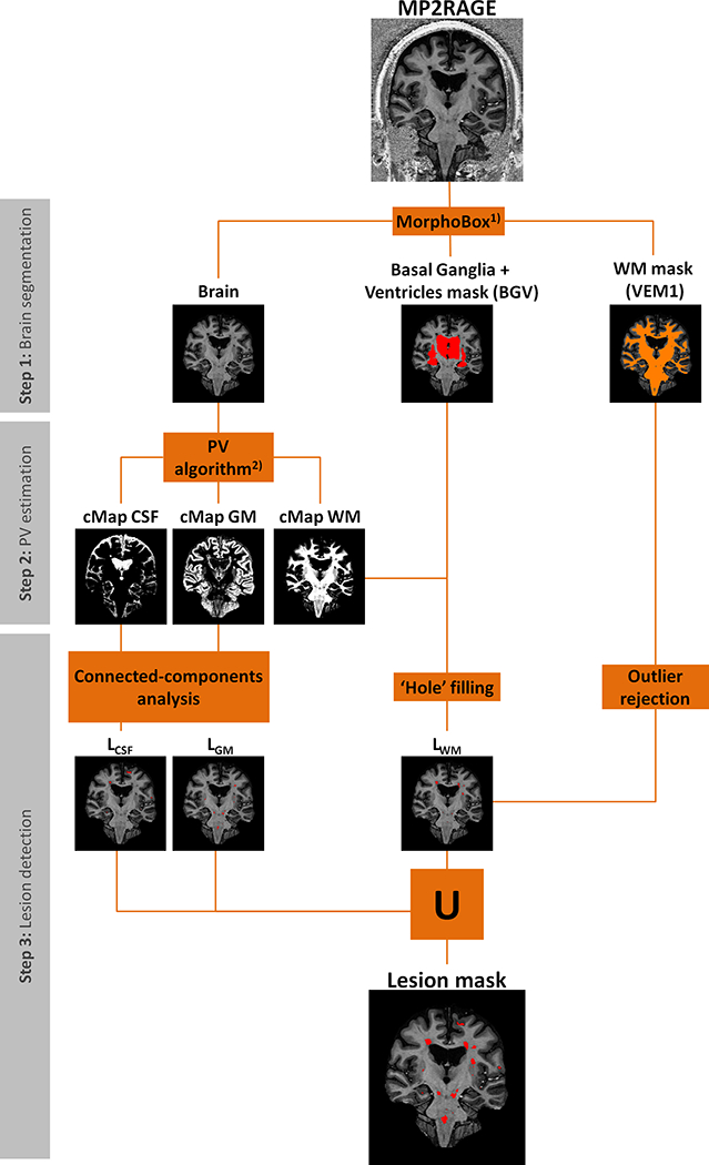
Figure 1 -.

Schematic diagram of the MSLAST method for lesion segmentation using 7T MP2RAGE, divided into three main steps: brain segmentation, partial volume (PV) estimation, and post-processing. VEM 1 is a template-based WM segmentation. cMap CSF, cMap GM, and cMap are concentration maps of white matter, gray matter, and cerebrospinal fluid, respectively. LCSF, LGM, and LWM are “pseudo” lesion masks computed from the cMaps of CSF, GM and WM, respectively. 1) - (25), 2) - (28).
