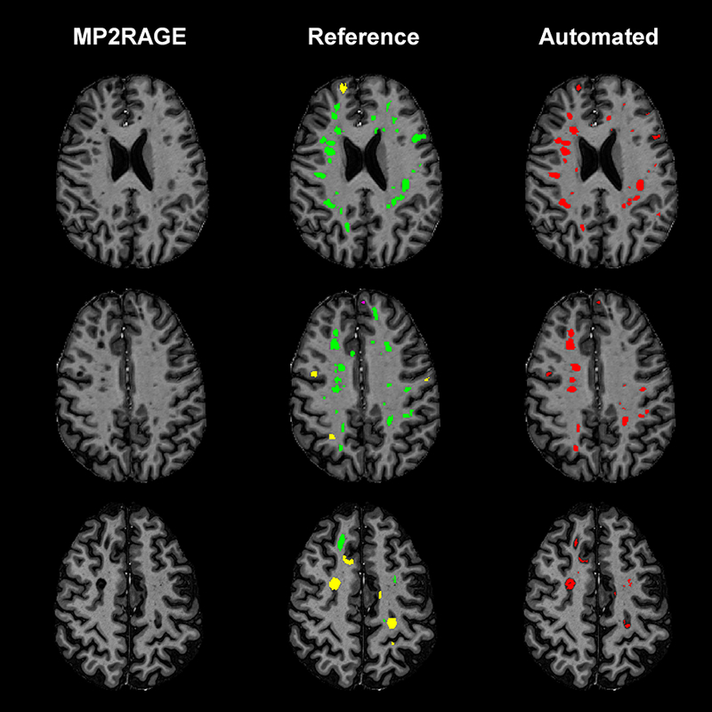
Figure 5 -.

Axial slices from three different MS patients showing the detection and segmentation results of MSLAST. From left to right: MP2RAGE skull-stripped image, manual segmentation, and automated segmentation. In the manual segmentations, white matter lesions are shown in green, leukocortical lesions in yellow, and intracortical lesions in pink.
