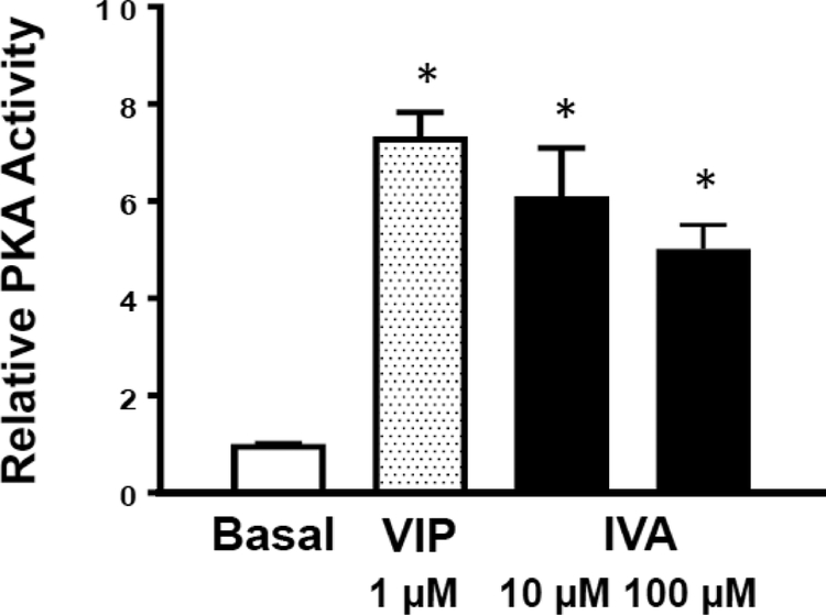
Figure 6: IVA-induced PKA activation in colonic muscle cells.
PKA activity in smooth muscle cells was measured by an in vitro kinase assay by the phosphorylation of PKA substrate kemptide using [32P]ATP. PKA activity was measured as described in the Methods and represented as relative PKA activity (ratio of counts per minute in the absence of added cAMP (stimulated activity) or in the presence of added cAMP (total activity)). Values are means±SD; * indicates P<0.05.

