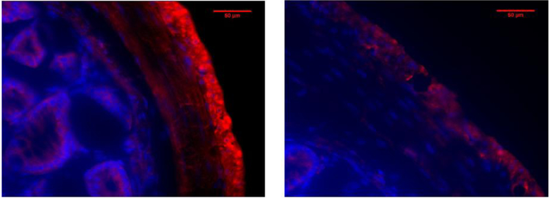
Figure 7: Fluorescent staining of mouse colon for Olfr558 (OR51E1).
Crossections (10 μm thick) of mouse colon demonstrate staining for Olfr558 (Red) in the longitudinal layers of smooth muscle, with some noticeable staining in the circular muscle and epithelium (A). Sections without primary antibody (B) did not show any noticeable fluorescence for the secondary antibody. Blue indicates nuclear DAPI staining. Magnification is 400X.

