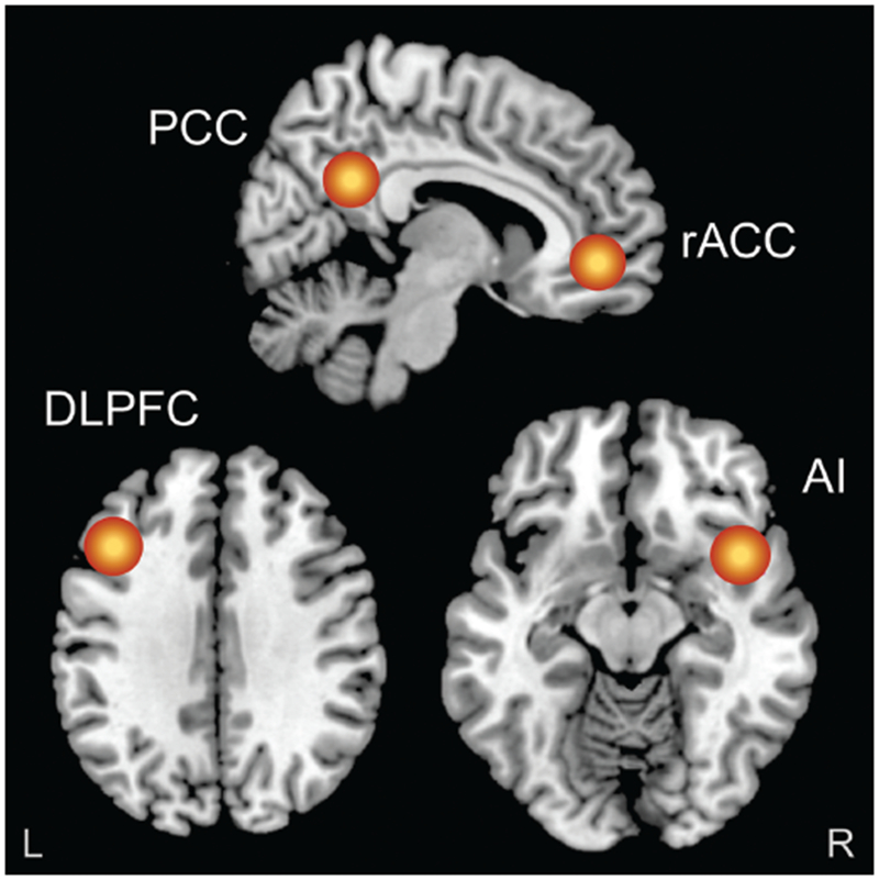
Figure 1.

Figure shows the regions of interest (10 mm radius) that were created in the rostral anterior cingulate cortex (rACC), posterior cingulate cortex (PCC), left dorsolateral prefrontal cortex (DLPFC) and right anterior insula (rAI). Resting-state functional connectivity was then computed (by means of lagged phase synchronization) between the rACC, and the PCC (default mode network), left DLPFC (frontoparietal network) and rAI (salience network), in both the theta (4.5-7 Hz) and beta (12.5-21 Hz) frequency bands. For the purposes of visualization, regions of interest shown here are displayed on a 2 × 2 × 2 Montreal Neurological Institute template brain (5 mm resolution is used for analyses in eLORETA).
