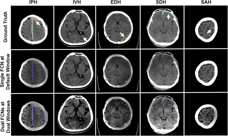
Fig. 11.
Examples of positively segmented CT images on third row using the dual FCN deep learning model, whereas they are predicted as negatives on the second row using a single FCN model trained on the default CT window setting. On the first-row images, the white arrow indicates ground truth of each hemorrhagic type

