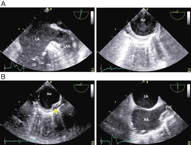
Figure 6.
(A) Dense SEC in the descending aorta (Ao) coincidentally found with LA SEC from a patient with nonvalvular atrial fibrillation. (B) Slight SEC in the descending aorta (Ao) with a mural plaque (arrow) observed from another patient with nonvalvular atrial fibrillation. Note that SEC in the right atrium (RA), swiftly flowing from the superior vena cava (SVC), is denser than SEC in the LA, indicating that some pathological factors that generate RA SEC might be involved in this patient.

 This work is licensed under a