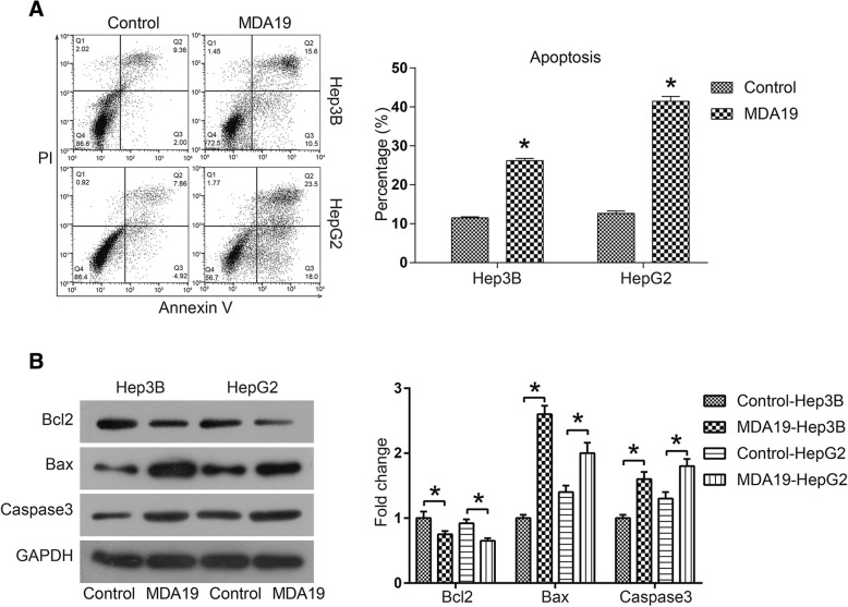
Fig. 2.
MDA19 induced apoptosis of HCC cells by activating mitochondrial-dependent apoptosis pathway. a Cell apoptosis of HCC cells treated with MDA19 (30 μM for Hep3B and 40 μM for HepG2) for 48 h was detected by a PI-AnnexinV-FITC assay and flow cytometry; The data were analyzed using FlowJo software. b Hep3B and HepG2 cells were treated with MDA19 for 48 h at 30 μM and 40 μM, respectively. Then the expression of apoptosis related proteins Bcl2, Bax and Caspase3 was detected by western blot and analyzed by Image J software. All experiments were performed at 3 times. *P < 0.05

