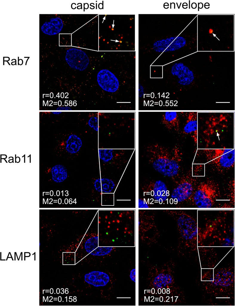
Fig. 12.

Co-localization between ZIKV and subcellular marker proteins in NH4Cl-treated Vero cells 20 min p.i. Confocal images of 50 mM NH4Cl ZIKV-infected Vero cells presenting co-localization between ZIKV capsid or envelope protein and Rab7, Rab11 and LAMP1 at 20 min p.i.. Rab7 – late endosomes marker protein, Rab11 – slow recycling endosomes marker protein, LAMP1 – lysosomes marker protein. ZIKV proteins are visualized in green, cellular proteins in red and nuclei are shown in blue. Co-localization coefficients for the representative presentations are indicated in the bottom left corners of the respective images; r – Pearson’s coefficient; M2 - Manders’ coefficient M2 (the virus overlapping with Rab7/Rab11/LAMP1). Scale bar = 10 μm
