Fig. 13.
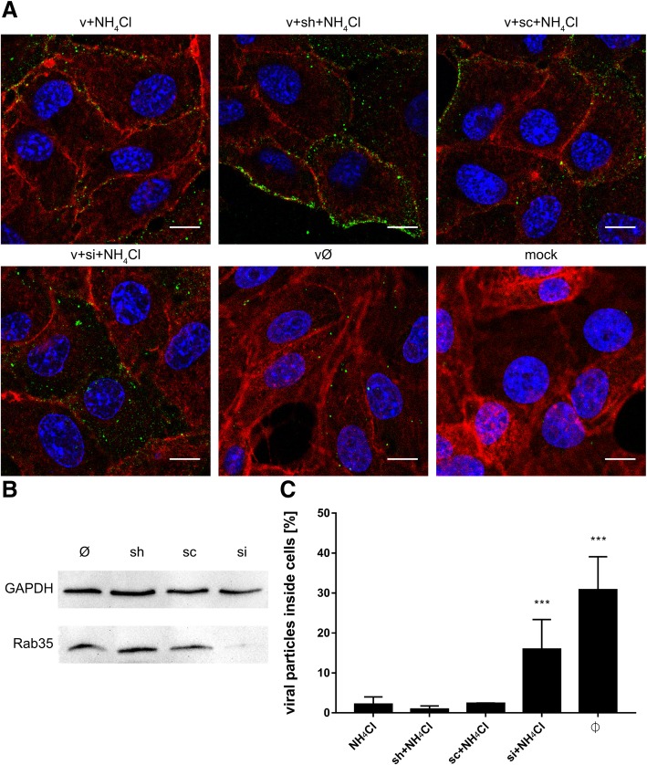
Subcellular localization of ZIKV in Rab35-depleted cells upon NH4Cl treatment. (a) Confocal analysis of ZIKV localization 1 h p.i. in NH4Cl-treated Vero cells. ZIKV envelope protein is visualized in green, actin cytoskeleton stained in red to show the cell boundaries and nuclei are shown in blue. Scale bar = 10 μm. (b) Western blot analysis of the efficiency of siRNA-dependent Rab35 silencing (Rab35 expression in Vero cells compared to GAPDH expression in these cells). M – BlueStar prestained protein marker; Ø – normal non-transfected Vero cells. (c) Graph representing the percent of ZIKV particles present inside cells related to the total number of ZIKV particles, assessed from confocal images of siRNA-transfected and all control Vero cells infected with ZIKV H/PF/2013 in the presence of 50 mM NH4Cl. v + NH4Cl - ZIKV-infected, NH4Cl-treated, non-transfected cells; v + sh + NH4Cl - ZIKV-infected, NH4Cl-treated, sham transfected cells; v + sc + NH4Cl - ZIKV-infected, NH4Cl-treated, scrambled siRNA transfected cells; v + si + NH4Cl - ZIKV-infected, NH4Cl-treated, Rab35-specific siRNA transfected cells; vØ - ZIKV-infected, NH4Cl-untreated, non-transfected cells; mock - mock-infected, NH4Cl-untreated, non-transfected cells. The data is presented as the mean ± SD. To determine the significance of differences between compared groups, single-factor analysis of variance (ANOVA) was applied. P values < 0.05 were considered significant. One asterisk (*) identifies adjusted P values between 0.01 and 0.05, two asterisks (**) identify adjusted P values between 0.01 and 0.001, three asterisks (***) identify adjusted P values between 0.001 and 0.0001

