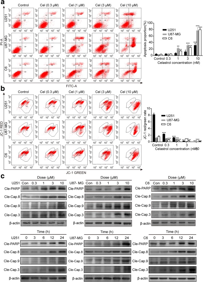
Fig. 3.
Celastrol induced intrinsic and extrinsic apoptosis in glioma cells. a Cells were treated with celastrol (3 μM) for 24 h, early and late apoptotic cells were analyzed using Annexin V-FITC/PI flow cytometry. The histograms indicate the proportion of apoptosis from three separate experiments. b The MMP was measured with the fluorescent mitochondrial probe JC-1 and assessed by flow cytometry. The histograms indicate the red/green fluorescence intensity ratio. c Cells were treated with various concentrations of celastrol for 24 h or incubated with celastrol (3 μM) for different durations. The apoptosis-related proteins cleaved PARP and Caspase-3, -8, -9 were analyzed by western blotting. β-actin was used as an internal control. Data are presented as the Mean ± SD (n = 3). *P < 0.05, **P < 0.01, ***P < 0.001, significantly different compared with the untreated control group

