Fig. 4.
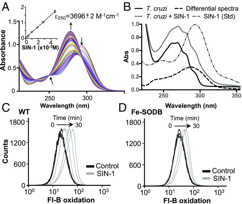
Fe-SODB limits SIN-1–derived peroxynitrite generation inside the parasite. (A) UV-visible spectra of 0.1 mM SIN-1 [in PBS, pH 7.4, containing 0.1 mM diethylenetriamine pentaacetic acid (DTPA)] recorded at 1-min intervals. The isosbestic point at 250 nm is shown. (Inset) Extinction coefficient (ε) of SIN-1 (0 to 5 mM) at 250 nm. (B) Parasites were preloaded, or not (control), with SIN-1, and proteins were precipitated. Spectra from control and SIN-1 parasites were recorded, and the differential spectra were obtained. A spectrum from SIN-1 (0.1 mM) is shown (Std). Absorbance at 250 nm was used to estimate intracellular SIN-1 concentration (ε250 = 3,696 M−1⋅cm−1). (C and D) Fl-B–preloaded epimastigotes (1 × 10⁸) from WT (C) and Fe-SODB parasites (D) were incubated at 28 °C in dPBS in the presence or absence of 0.1 mM SIN-1. Intracellular fluorescence was analyzed by flow cytometry; arrows indicate fluorescence peak movement.

