Fig. 5.
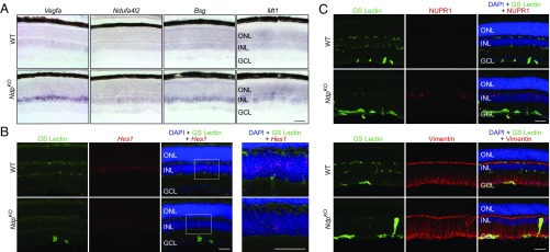
Histochemical assessment of transcripts and proteins validates differentially regulated Muller glia genes in WT and NdpKO retinas. (A) Abundance changes for four transcripts that are up-regulated in NdpKO compared with WT retinas. In situ hybridization shows increased abundance in the INL in the NdpKO retina. (Scale bar: 50 μm.) (B) Fluorescent Hes1 in situ hybridization shows decreased abundance in the INL in the NdpKO retina. The boxed region encompassing the INL is enlarged in Right. (Scale bars: 50 μm.) (C, Upper) Immunofluorescence of retina cross-sections showing blood vessels (GS Lectin; green), Nupr1 (red), and nuclei (DAPI; blue). NUPR1 is present in scattered Muller glial cell bodies in the NdpKO retina but is undetectable in WT retinas. (C, Lower) Immunofluorescence of retina cross-sections showing blood vessels (GS Lectin; green), Vimentin (Vim; red), and nuclei (DAPI; blue). Vimentin is modestly elevated in the NdpKO retina compared with the WT retina. GCL, ganglion cell layer; ONL, outer nuclear layer. (Scale bars: 50 μm.)

