Fig. 3.
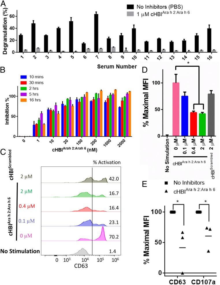
cHBIs inhibit degranulation induced by crude peanut extract in clinical patient samples. (A) cHBIArah2:Arah6 inhibits degranulation induced by crude peanut extract. A total of 1 µM cHBIArah2:Arah6 inhibited degranulation induced by crude peanut extract in RBL-SX38 cells primed with 16 different patient sera. Note that due to differences in patient sensitivity to crude peanut extract, patient samples were challenged with varying amount of crude peanut extract after cHBI incubation. Patients 1, 2, and 4 were challenged with 1 μg/mL peanut extract, patients 3 and 7 with 100 ng/mL, patient 5 with 10 ng/mL, and all others with 1 ng/mL PBS, used as a control. (B) RBL-SX38 cells primed with patient serum (serum 4) were incubated with varying concentration of cHBIArah2:Arah6 for varying time points (10 min, 30 min, 2 h, 5 h, and 16 h), washed, and then challenged with 100 ng/mL of crude peanut extract. (C) cHBIArah2:Arah6 inhibits degranulation in BAT. BAT data for whole blood incubated with 0 μM (pink), 0.1 µM (blue), 0.4 µM (red), or 2 µM (green) cHBIArah2:Arah6 and then challenged with 100 ng/mL crude peanut extract are shown. Flow cytometry plots are shown with fluorescence intensity on the x axis and number of basophils on the y axis. Controls include blood incubated without crude peanut extract (no treatment), and blood incubated with 2 µM of a 1:1 mixture of cHBIs presenting Scrambled sequences of Ara h 2 epitope 5 and Ara h 6 epitope 5, respectively (cHBIScrambled). Basophils were considered CD63 positive if fluorescence intensity was greater than the threshold of 2 × 10⁴ (black line). Percent activated basophils is shown on Right side of plot. (D) Percent maximal MFI of flow cytometry plots from D is shown, calculated as percentage between the “0 µM” and “no stimulation” MFI values. Error bars indicate ± SD of triplicate experiments. (E) Three additional patients were assessed using BAT assay. Whole blood from three patients was incubated with (triangles) or without (squares) 1 μM cHBIArah2:Arah6 and then challenged with 100 ng/mL of crude peanut extract. Percent maximal MFI was similarly calculated as in D. Each point represents an average of three technical replicates of each patient; horizontal lines represent mean of all three patients. *P < 0.05.

