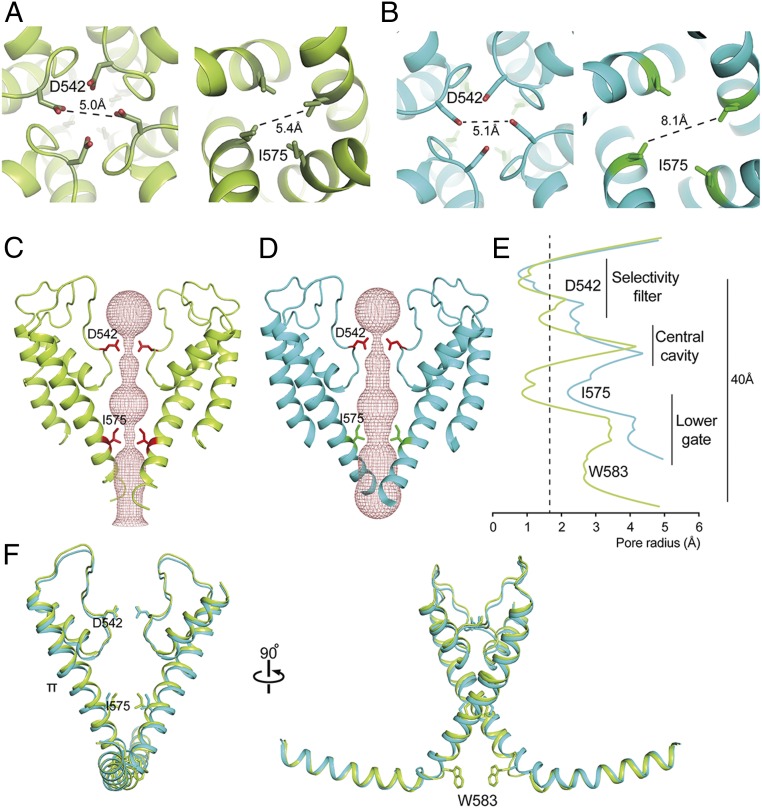
Fig. 3.
Pore domain characteristics of TRPV5. (A and B) Top (Left) and bottom (Right) view of TRPV5 full-length (A) and TRPV5 W583A (B). Side chains are shown for two constricting residues in the selectivity filter and lower part of the pore, D542 and I575. (C and D) The ion permeation pathway of closed TRPV5 (C; yellow) and open TRPV5 W583A (D; cyan) is shown as a ribbon diagram with the solvent-accessible space depicted as a pink mesh. Only two subunits are shown for clarity, with side chains of the restricting residues D542 and I575. (E) Diagram of the pore radius calculated with HOLE shown for TRPV5 (yellow) and TRPV5 W583A (cyan). The dotted line indicates the radius of a hydrated calcium ion. (F) Comparison of the TRPV5 (yellow) and TRPV5 W583A (cyan) pore domains with the TRP helix attached. Key residues in the pore domain are depicted.

