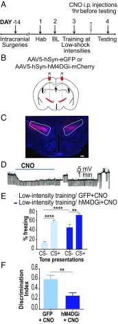
Fig. 2.
Decreasing cellular activity in the ZI results in fear generalization. (A) Experimental protocol for chemogenetic inhibition. Two weeks after intracranial injection of the control or DREADD virus, animals were conditioned to low-threat intensities. The next day, CNO was administered i.p. 1 h before testing fear generalization. BL, baseline; Hab, habituation. (B) Wild-type animals were injected with either the control virus (AAV5-hSyn-EGFP) or inhibitory DREADDs (AAV5-hSyn-hM4DGi-mCherry) at −1.52 mm posterior to the bregma. (C) Representative image of the ZI targeted with intracranial infusions of DREADD-expressing mCherry viruses (in red) and Hoechst-stained nuclei (in blue). (Scale bar: 100 μm.) (D) Patch-clamp recording of hSyn-hM4DGi-mCherry–expressing cells in the ZI showing membrane hyperpolarization during CNO exposure. (E) Chemogenetic inhibition of the ZI (hM4DGi+CNO) resulted in a significant increase in fear response to CS− compared with controls (GFP+CNO). (F) Chemogenetic inhibition of the ZI (hM4DGi+CNO) resulted in an impaired ability to discriminate between the CS+ and the CS−. **P < 0.01; ****P < 0.0001. Data are represented as mean ± SEM.

