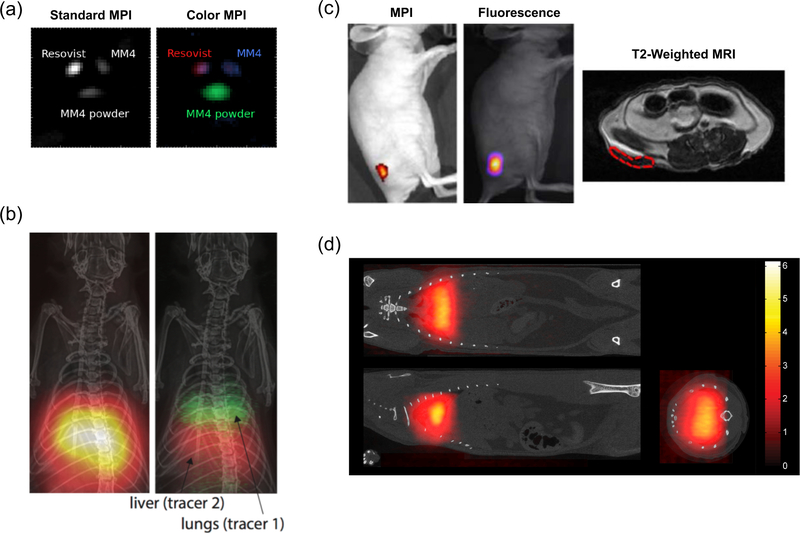
Figure 4. Recent progress in tracer technologies.
(a) In vitro tri-color MPI. Three different MPI tracers are indistinguishable in a standard MPI reconstruction algorithm, but can be distinguished after applying a multi-color reconstruction algorithm. Adapted with permission from51 under the Creative Commons Attribution 3.0 license (http://creativecommons.org/licenses/by/3.0). (b) In vivo dual-color MPI. Rat lung and liver are targeted with two nanoparticles with different relaxation behavior. In standard MPI, the organs are indistinguishable, but after the colorizing algorithm the organs can be distinguished based on the relaxation behavior of the SPIOs within. Image courtesy of Daniel Hensley. (c) Multi-modal Janus iron oxide MPI tracers. SPIO tracers can be designed for multi-modality imaging. Mice were subcutaneously implanted with nanoparticle-labeled cells and imaged under MPI, fluorescence and T2-weighted MRI. Adapted with permission from53•. Copyright 2017 American Chemical Society. (d) Lung perfusion imaging with MAA-SPIO. Large macroaggregated albumin conjugated to SPIOs are biomechanically trapped in the rat lung, allowing imaging of blood perfusion through the lungs11. Institute of Physics and Engineering in Medicine. Adapted with permission of IOP Publishing. All rights reserved.

