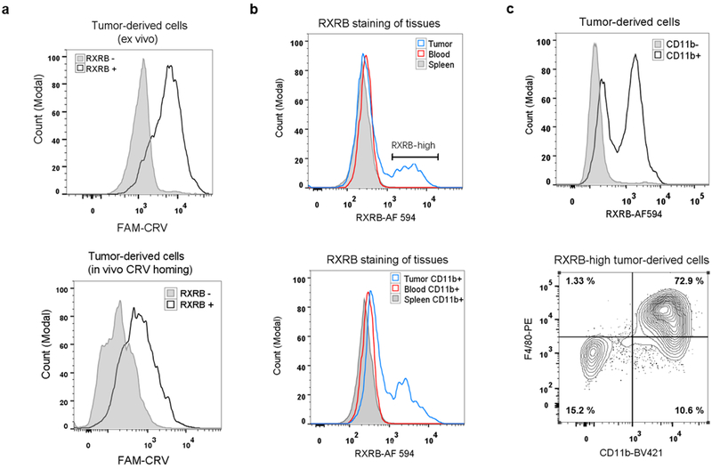
Fig. 5.
Validation of RXRB as the CRV receptor in TAMs. (a) Flow cytometry histograms show preferential binding of FAM-CRV to RXRB+ over RXRB− 4T1 tumor cells. Top: 4T1 tumor cells incubated with FAM-CRV and anti-RXRB antibody ex vivo. Bottom: After in vivo FAM-CRV 1-h homing, dissociated 4T1 tumor cells were incubated with anti-RXRB antibody. (b) Flow cytometry histograms show RXRB expression in single cells of the indicated tissues. Top: RXRB expression in tumor, spleen, and blood whole cell populations. Bottom: RXRB expression in tumor, spleen, and blood CD11b+ cells. AF 594: Alexa Fluor 594. (c) Flow cytometry analysis showing high RXRB expression on tumor macrophages. Top: Comparison of RXRB expression on CD11b+ and CD11b− cells in tumor-derived cell suspension. Bottom: phenotypic analysis of RXRB-high cells from 4T1 tumors for the indicated macrophage markers. BV421: Brilliant Violet 421. PE: phycoerythrin. All experiments were performed on individual samples from three animals per group, and representative images are shown.

