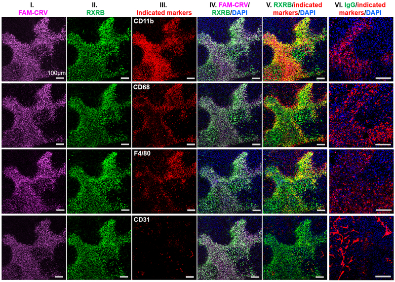
Fig. 6.
Co-localization of systemically administered FAM-CRV, rabbit polyclonal anti-RXRB antibody, and macrophages in 4T1 tumors. After in vivo homing of FAM-CRV and anti-RXRB antibody (or control IgG) in mice bearing 4T1 breast tumors, IF staining was performed with anti-FITC (column I, magenta), anti-rabbit IgG secondary antibody for RXRB (column II, green), other indicated antibodies (column III, red) and DAPI (blue). Column IV shows merged images of FAM-CRV, anti-RXRB antibody, and DAPI staining. Column V shows merged images of anti-RXRB antibody with the indicated markers and DAPI. Column VI represents merged images of control IgG, the indicated markers, and DAPI staining of tumors from mice that received the control IgG. Scale bar: 100 μm. The experiments were performed using three animals per group (anti-RXRB or IgG), and representative images are shown.

