Figure 1.

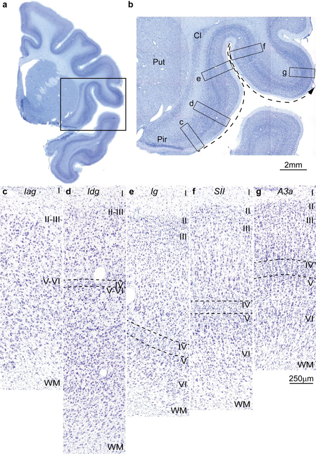
Cortical types and laminar gradients of differentiation in the insula and adjacent frontal cortex of the rhesus monkey (Nissl staining). a, Coronal section of the rhesus monkey brain at the level of the frontotemporal junction stained for Nissl. b, Closer view of the insula and the frontal operculum, shows the levels of photomicrographs in c-g; the dashed arrow shows the increasing trend of laminar differentiation. c, The agranular insula (Iag) is next to the primary olfactory cortex (Pir in b) and lacks layer IV. d, Dysgranular insula (Idg) has a rudimentary layer IV (dashed lines). e, Layer IV (dashed lines) is better developed in the adjacent granular insula (Ig). f, The secondary somatosensory area (SII) has a denser layer IV (dashed lines) and better differentiation of the other layers than the granular insula. g, Area 3a, which is part of the primary somatosensory cortex, has thicker and denser layer IV (dashed lines) and its superficial layers are denser than in SII. Abbreviations: A3a: area 3a, Cl: claustrum; Iag: insula agranular; Idg: insula dysgranular; Ig: insula granular; Pir: piriform cortex in the primary olfactory cortex; Put: putamen; SII: secondary somatosensory area; WM: white matter. Roman numerals indicate cortical layers. Calibration bar in g applies to c-g.
