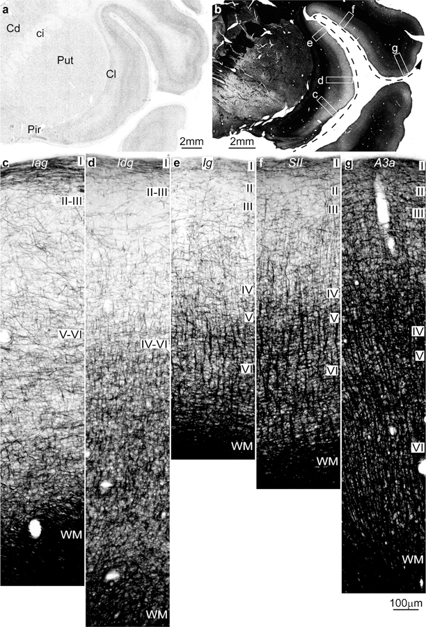
Figure 2.

Intracortical myelin varies systematically in parallel with laminar differentiation in the insula and adjacent frontal cortex in the rhesus monkey (Gallyas staining). a, Coronal section of the rhesus monkey brain at the level of the frontotemporal junction stained for Nissl shows the insula and the frontal operculum. b, Adjacent section to a is stained for myelin (black) and shows the levels of photomicrographs c-g; the dashed arrow shows the trend of increasing laminar differentiation. c, The agranular insula (Iag) is next to the primary olfactory cortex (Pir in a) and has comparatively fewer myelinated axons than the adjacent area. d, Dysgranular insula (Idg) is slightly better myelinated. e, The granular insula (Ig) has more myelinated axons organized into vertical bundles than Iag and Idg. f, The secondary somatosensory area (SII) has more myelinated axons than the insular areas with well-developed vertical bundles in the deep layers and abundant small horizontal axons in the middle layers. g, Area 3a (A3a) in the primary somatosensory cortex has more myelinated axons than SII in all layers. Abbreviations: A3a: area 3a, Cd: caudate; ci: internal capsule; Cl: claustrum; Iag: insula agranular; Idg: insula dysgranular; Ig: insula granular; Pir: piriform cortex in the primary olfactory cortex; Put: putamen; SII: secondary somatosensory area; WM: white matter. Roman numerals indicate cortical layers. Calibration bar in g applies to c-g.
