Figure 3.

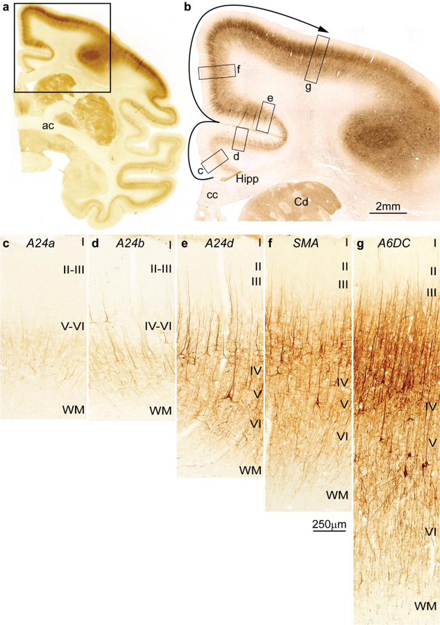
Cortical types and laminar gradients of differentiation in the cingulate and frontal cortex of the rhesus monkey (SMI-32 staining). a, Coronal section of the rhesus monkey brain at the level of the anterior commissure (ac) stained for the nonphosphorylated neurofilament protein SMI-32. b, Higher magnification shows the cingulate and dorsal frontal cortex and the levels of photomicrographs c-g; the solid arrow shows the trend of increasing laminar differentiation. c, Agranular area 24a (A24a) in the cingulate cortex is next to the anterior extension of the hippocampal formation (Hipp in b) and has few SMI-32 labeled neurons restricted to the deep layers. d, Area 24b (A24b) in the lower bank of the cingulate sulcus has few labeled SMI-32 neurons in the deep layers and some at the bottom of layer III. e, Area 24d (A24d) in the upper bank of the cingulate sulcus has more SMI-32 neurons than the other cingulate areas in the deep layers and in layer III, demarcating a band of unstained tissue that corresponds to layer IV. f, The supplementary motor area (SMA) and (g) premotor area 6 dorsocaudal (A6DC) have more neurons labeled for SMI-32 than the cingulate areas; in both SMA and A6DC layer IV stands out as a band of unlabeled tissue between the bottom of layer III and the upper part of layer V. Abbreviations: A24a: cingulate area 24a; A24b: cingulate area 24b; A24d: cingulate area 24d; A6DC: premotor area 6 dorsocaudal; ac: anterior commissure; cc: corpus callosum; Cd: caudate; Hipp: anterior extension of the hippocampal formation; SMA: supplementary motor area; WM: white matter. Roman numerals indicate cortical layers. Calibration bar in f applies to c-g.
