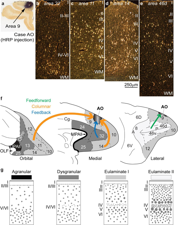
Figure 5.

Examples of connections predicted by the relational Structural Model. a, Injection of the neural tracer horseradish peroxidase (HRP) in area 9 of the dorsolateral prefrontal cortex in a rhesus monkey brain. b, Retrogradely labeled neurons in anterior cingulate area 32 are concentrated in the deep layers V-VI, typical of a feedback pattern of connections originating from an area with a simpler laminar structure (area 32) and directed to an area with a more elaborate laminar structure (area 9). c-d, In area 11 (c) and in area 14 (d) there are labeled neurons in superficial layers II-III and in the deep layers V-VI, typical of a columnar pattern of connections between areas of a comparable type; note absence of labeled neurons in layer IV, which do not project out of the cortex. e, Retrogradely labeled neurons in the dorsal part of area 46d are concentrated in the superficial layers II-III, typical of a predominant feedforward pattern of connections, originating from an area with a more elaborate architecture (area 46d) to an area with comparatively simpler architecture (area 9). f, Sketches of the prefrontal cortex of the rhesus monkey show the orbital (left), medial (center), and lateral (right) surfaces; agranular areas are shown in black, dysgranular areas in dark grey, and eulaminate areas are depicted progressively with lighter grey. Arrow color indicates type of connection; arrow thickness indicates relative strength of pathways; thicker arrows suggest more robust connections. g, Cortical types (agranular, dysgranular, eulaminate I, and eulaminate II) are represented in four sketches. Abbreviations: HRP: horseradish peroxidase; MPAll: medial periallocortex; OLF: primary olfactory cortex; OPAll: orbital periallocortex; OPro: orbital proisocortex. Arabic numerals correspond to prefrontal cortical areas according to Barbas and Pandya (1989). Roman numerals indicate cortical layers. Calibration bar in d applies to b-e.
