Figure 9.

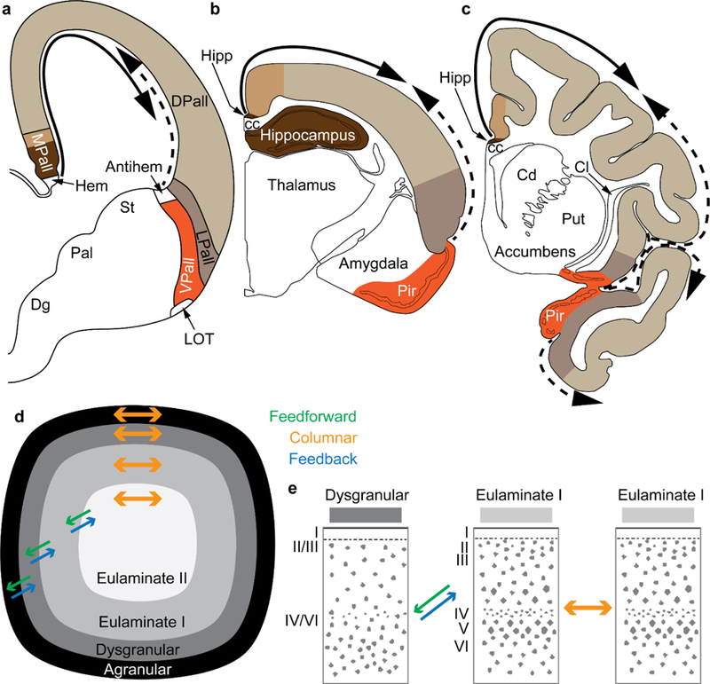
Two organizers for the expansion of the neocortex in development and evolution: the hem and antihem. a, Sketch of the mammalian telencephalon in development in coronal section shows the pallial (cortical) sectors [medial pallium (MPall), dorsal pallium (DPall), lateral pallium (LPall), and ventral pallium (VPall)], the hem, and the antihem according to the terminology of Subramanian et al. (2009), Montiel and Aboitiz (2015), and Puelles (2017) with the addition of the distinction of two parts on the MPall sector corresponding to allocortex (hippocampus) and periallocortex (agranular/dysgranular cingulate areas) in rats (b) and primates (c) based on architectonic analysis of these species in adults. The hem, found next to the roof plate, and the antihem, found in the corticostriatal junction, are secondary organizers that secrete morphogen proteins that form two overlapping gradients (solid and dashed arrows). b, Sketch of the rat adult brain in a coronal section; the adult derivatives of the developmental pallial sectors are colored as in a. The solid arrow shows the trend of laminar differentiation traced to the ancestral hippocampal cortex; the dashed arrow shows the trend of laminar differentiation traced to the ancestral olfactory cortex (Pir). c, Sketch of the adult rhesus monkey brain in coronal section; the adult derivatives of the developmental pallial sectors are tinted as in a. The solid arrow shows the dorsal trend of laminar differentiation traced to the ancestral hippocampal cortex; the dashed arrow shows the ventral trend of laminar differentiation traced to the ancestral olfactory cortex Pir). Note that DPall derivatives are more expansive than in the rat and extend to dorsal and ventral regions. d, Schematic of the primate cerebral cortex shows the arrangement of cortical types in rings. Laminar differentiation progresses from the outer or basal (black and dark grey) to the inner rings (lighter shades of grey). The edge of the cortex (black and dark grey) is actually thin compared to the greatly expanded eulaminate areas in the center. Cortical areas have stronger connections with other areas in the same ring and display columnar patterns of connections (orange arrows). Connections between areas in different rings (i.e., of different cortical type) are less strong than connections within the same ring and display feedback (blue arrows) and feedforward (green arrows) laminar patterns of connections. e, according to the Structural Model (Barbas and Rempel-Clower 1997), the laminar pattern of connections is related to the cortical type difference of the connected areas. Pathways form dysgranular to eulaminate areas are feedback (blue arrow); pathways from eulaminate to dysgranular are feedforward (green arrow); pathways between areas of comparable cortical type are columnar (orange arrow). Abbreviations: cc: corpus callosum; Cd: caudate; Cl: claustrum; Dg: Subpallial diagonal domain; DPall: dorsal pallium; Hipp: anterior extension of the hippocampal formation; LOT: lateral olfactory tract; LPall: lateral pallium; MPall: medial pallium; Pal: pallidum; Pir: piriform cortex in the primary olfactory cortex; Put: putamen; St: striatum; VPall: ventral pallium. Roman numerals indicate cortical layers.
