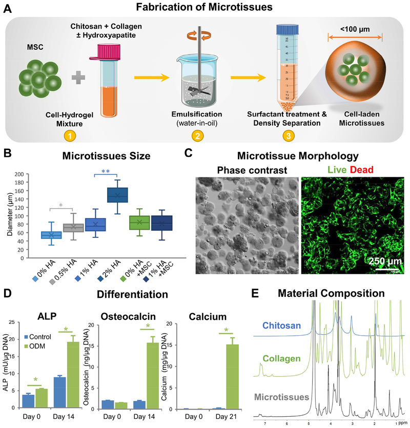
Figure 1 -. Fabrication and characterization of chitosan-collagen microtissues.
A) Schematic of the water-in-oil emulsification process used to embed MSC within CHI-COL composite microtissues. B) Size distribution of CHI-COL microtissues as a function of HA content and cell loading. C) Phase contrast (Day 0) and fluorescence images (Day 14) showing the morphology of microtissues and vital staining (green) of embedded MSC, respectively. D) Expression of osteogenic markers by microtissues cultured in control and osteogenic medium (ODM) in vitro. E) 1H-NMR spectra of the microtissue matrix materials, demonstrating the presence of collagen and chitosan.

