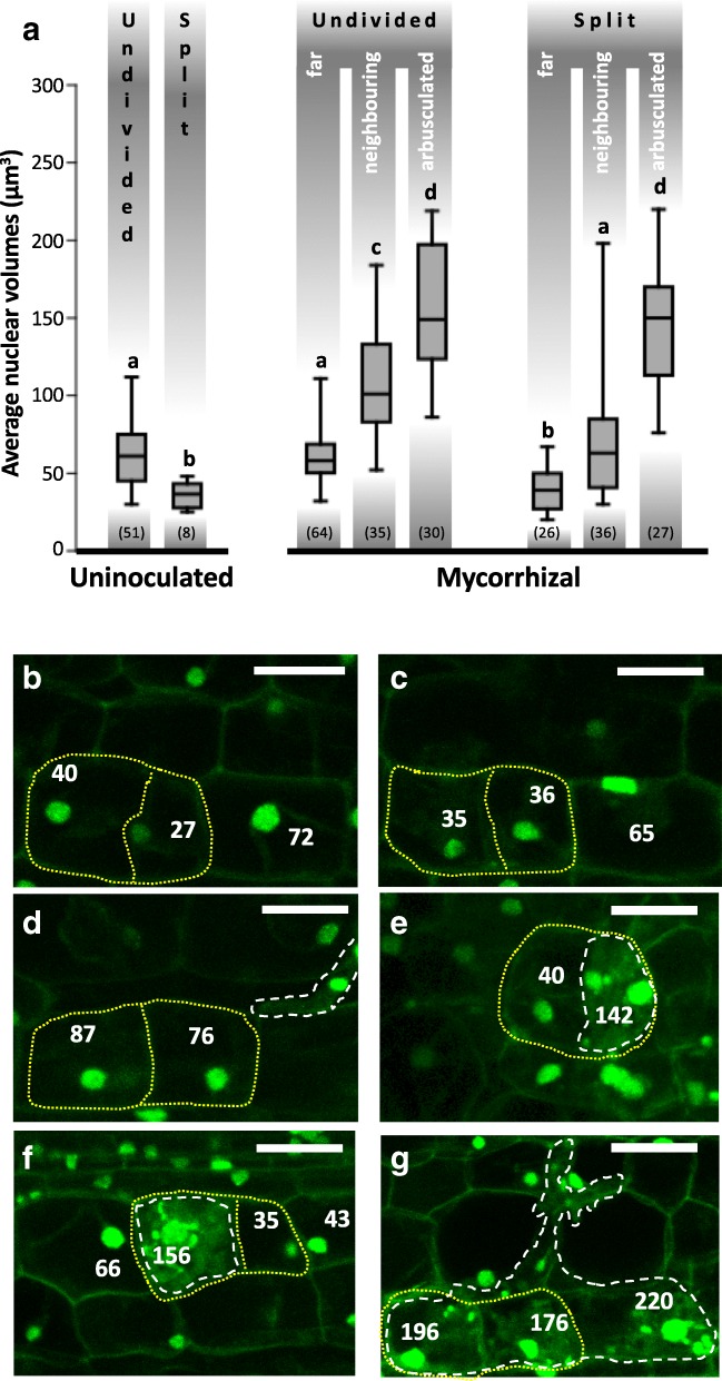
Fig. 5.
Increase in nuclear volumes follows cortical cell divisions throughout root colonization. a Average nuclear volume in non-divided and divided (here referred as split cells) cortical cells from uninoculated and mycorrhizal ROC segments of wild-type M. truncatula. For a more detailed representation of nuclear changes in wild-type mycorrhizal root cortex, we discriminated undivided and split cells in arbusculated from neighbouring and far uncolonized divided cells. Quantitative analysis was done on a dataset of 10 z-stacks from different uninoculated and mycorrhizal root segments. The graph shows a significant increase in average nuclear volume of arbusculated and uncolonized neighboring corticals in both undivided and split cells of mycorrhizal roots. The average nuclear volume of undivided corticals in uninoculated samples is comparable with that of far undivided cells of mycorrhizal roots. Also uncolonized split cells of uninoculated (split cells) and mycorrhizal (far split cells) sections show comparable average nuclear volumes. Bars represent standard deviations; letters indicate statstically significant differences based on Kruskal-Wallis non-parametric analysis of variance followed by Dunn’s post-hoc test (using Bonferroni-corrected p-values) for pairwise multiple comparisons (P < 0.05). The numbers in parentheses indicate the number of measurements for each experimental condition
The figure presents a collection of representative z-stack projections of progressive steps in wild-type M. truncatula ROC colonization by G. margarita of cortical divided cells. Numbers indicate the volume of each nucleus in μm3. Panel (b) shows cortical divided cells (yellow dashed outline) in uninoculated root cortex with nuclei belonging to volume class I (20–45 μm3), as defined by Sturges’ rule. Panel (c) shows the same feature in split cells from inoculated roots far from the fungal penetration units. In (d), an Intraradical hypha (white dashed outline) colonizing the cortex is shown, with a couple of split cells containing class III nuclei (70–95 μm3), suggesting the occurrence of ectopic cell division (Russo et al., 2018) and endoreduplication (Carotenuto et al., 2019) before cell penetration. Panels (e) and (f) present early infection units, with couples of split cortical cells where only one cell is arbusculated. Such colonized cells display increasingly enlarged nuclei, with sizes reaching class V (120–145 μm3) in panel (e) and VI (145–170 μm3) in panel (f). More advanced infection units are shown in panel (g): large nuclei - class VII (170–195 μm3) and VIII (195–220 μm3) - are present in both split and undivided arbusculated cells. Bars = 25 μm.

