Figure 1.
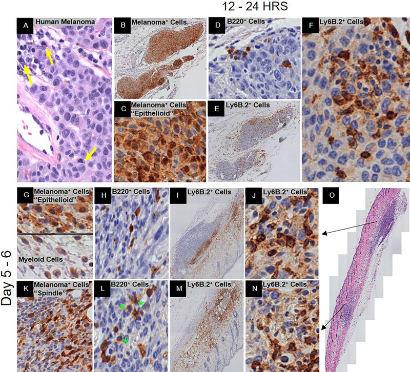
Spontaneous immunosurveillance. Representative histology is shown at low and/or high magnification. (A) H&E of human metastatic melanoma with plasma cell infiltration. Yellow arrows denote plasma cells. (B – O) C57BL/6J mice post-YUMMER1.7 implantation. At 12 – 24 hrs., (B, C) GFP+ melanoma cells (nucleus+cytoplasmic+), (D) B220+ B cells, and (E, F) Ly6B.2+ neutrophils. At 5–6 days, (G, K) GFP+ melanoma cells (nucleus+cytoplasmic+) and antigen presenting cells (nucleus−/lowcytoplasmic+), (H, L) B220+ B cells (green stars denote plasmablasts and/or plasma cells), (I, J, M, N) Ly6B.2+ neutrophils, and (O) H&E of biopsy. Black arrows depict the area of the biopsy specimen that the row represents.

