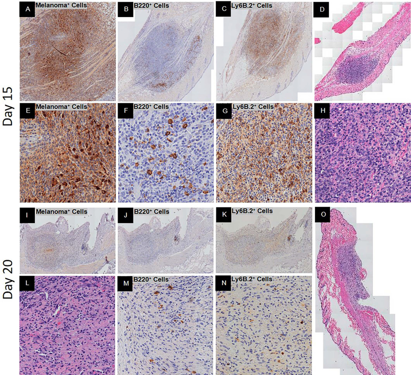
Figure 3.
Peak spontaneous adaptive immune response and complete spontaneous regression post-YUMMER1.7 implantation in C57BL/6J mice. Representative histology at low and/or high magnification. At 15 days, (A, E) GFP+ melanoma cells (nucleus+cytoplasmic+), (B, F) B220+ B cells with signs of fragmentation, (C, G) Ly6B.2+ neutrophils, and (D, H) H&E of biopsy. At 20 days, (I, L) GFP+ melanoma cells (nucleus+cytoplasmic+), (J, M) B220+ B cells, (K, N) Ly6B.2+ neutrophils, and (O) H&E of biopsy.

