Figure 1.

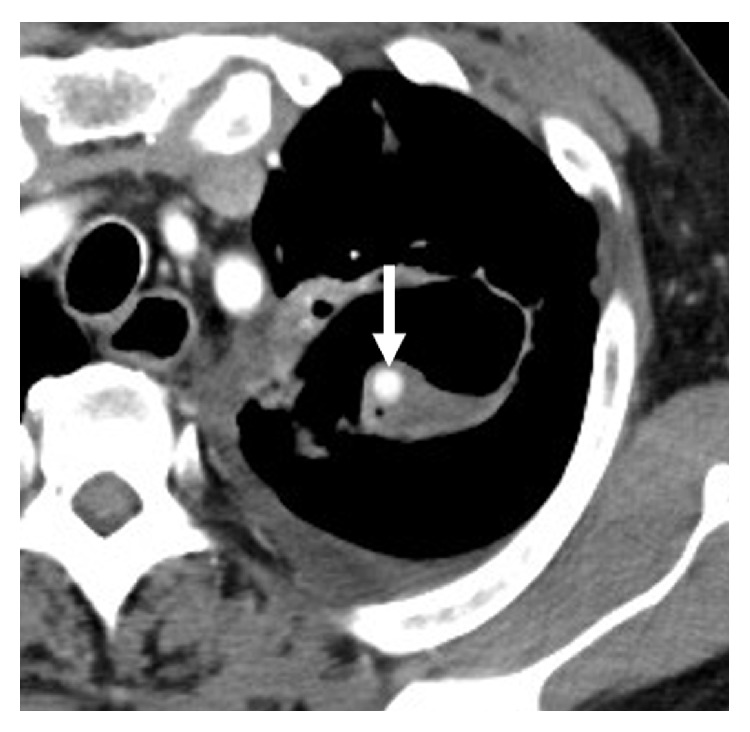
Contrast-enhanced axial computed tomography image. There is a 7 mm round aneurysm (arrow) within a cavitary lesion in the left upper lobe of the lung.

Contrast-enhanced axial computed tomography image. There is a 7 mm round aneurysm (arrow) within a cavitary lesion in the left upper lobe of the lung.