
Figure 2.
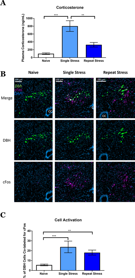
Immediate response of corticosterone and neuronal activation to restraint stress. A) Plasma corticosterone (ng/mL) from naïve, single stress, and repeat stress mice. Mean±SEM; n = 5–6 mice/group. One-way ANOVA (F = 15.07, p < 0.001). B) Nucleus of the solitary tract double fluorescent in situ hybridization showing DBH (green), cFos (lilac), and DAPI (blue) in stress naïve, single stress, and repeat stress mice. CC = central canal. C) Percent of DBH+ neurons that were colabeled for cFos. Mean±SEM; n =4–8 mice/group. One-way ANOVA (F = 11.31, p < 0.001). **p<0.05, *** p<0.005.