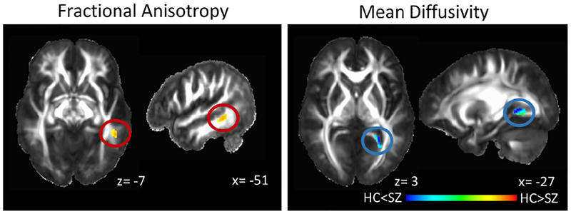
Figure 2:
White matter microstructural integrity abnormalities in never-treated and currently unmedicated patients with schizophrenia (SZ) compared to healthy controls (HC). Fractional anisotropy is decreased in the left medial temporal white matter (cluster extent: 123 voxels, peak: x= −51; y= −44; z= −7; α< .05) and mean diffusivity is increased in the fusiform/ lingual gyrus white matter extending to the hippocampal part of the cingulum (cluster extent 185 voxels, peak: x= −27; y= −49; z= 2; α< .04) in SZ compared to HC. Clusters are projected on the IIT2 white matter atlas template. Numbers adjacent to slices indicate x, y, and z coordinates in Talairach convention. Left= Right. Color bar indicates z scores.

