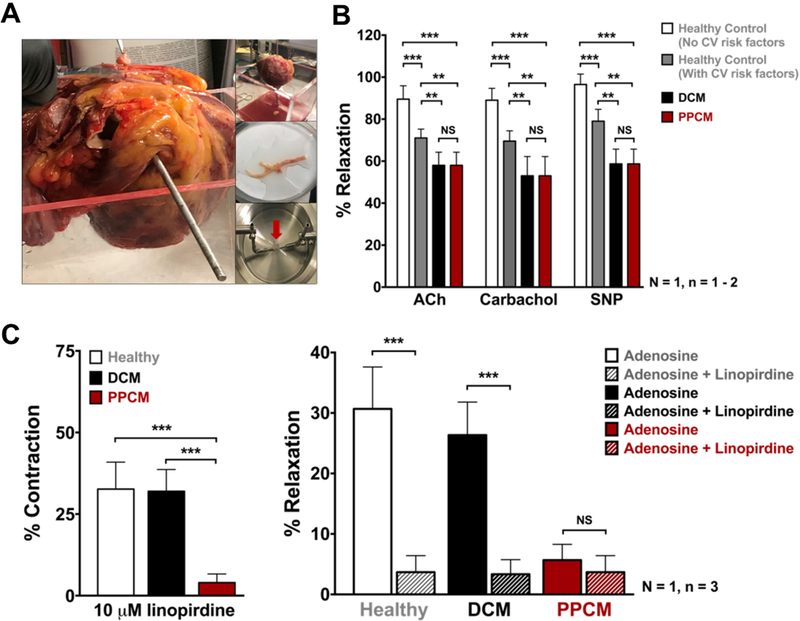
Figure 2. Vascular reactivity in PPCM heart.
(A) Explanted PPCM heart, dissected coronary arteries and wire-myograph. (B) Endothelium function in PPCM heart. Isometric tension recordings of relaxation to Acetylcholine (10μM), Carbachol (10μM), and NO donor SNP (3μM) upon pre-constriction with U46619. (C) Tension recordings of PPCM LAD segments showing less contraction at basal tone upon application of 10μM linopirdine when compared to DCM and healthy control (left panel); impaired adenosine response in PPCM or in presence of 10μM linopirdine (right panel). Statistical analyses were done on three segments (n=3) from one coronary vessel per patient (N=1) using two-way ANOVA. *p< 0.5, **p< 0.05, ***p< 0.01.

