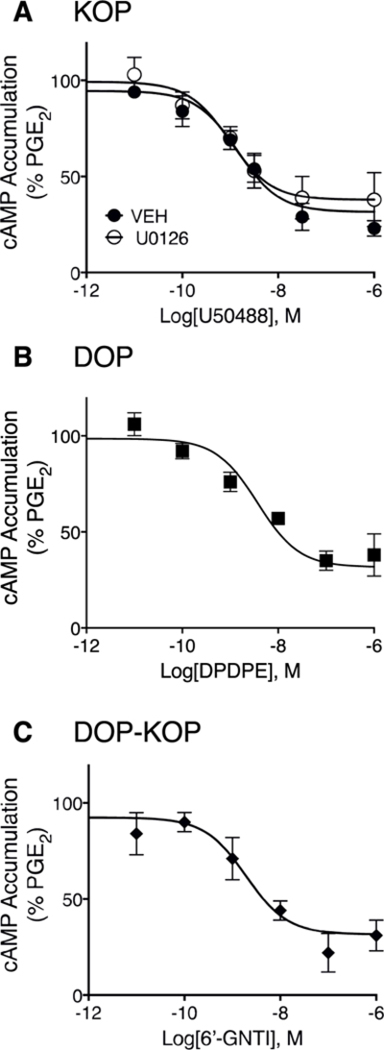
Fig. 3. Concentration response curves for U50488-, DPDPE- or 6’-GNTI-mediated inhibition of PGE2-stimulated cAMP accumulation in peripheral sensory neuron cultures.

Cells were incubated with BK (10 μM) for 15 min followed by further incubation with the indicated concentrations of either U50488 (A) DPDPE (B), or 6’-GNTI (C) along with PGE2 (1 μM) for 15 min. Cellular cAMP levels were measured with radioimmunoassay. Data are expressed as the percentage of PGE2-stimulated cAMP levels and are the mean ± SEM of 4 separate experiments. When not visible, error bars are contained within the symbol. Concentration-response curves were analyzed with non-linear regression to determine EC50 and Emax values that are provided in the text. As shown in panel A, U50488-mediated inhibition of PGE2-stimulated cAMP levels was not altered in cells pretreated with the MEK inhibitor, U0126 (10 μM); F(1,42) = 1.764, P = 0.19.
