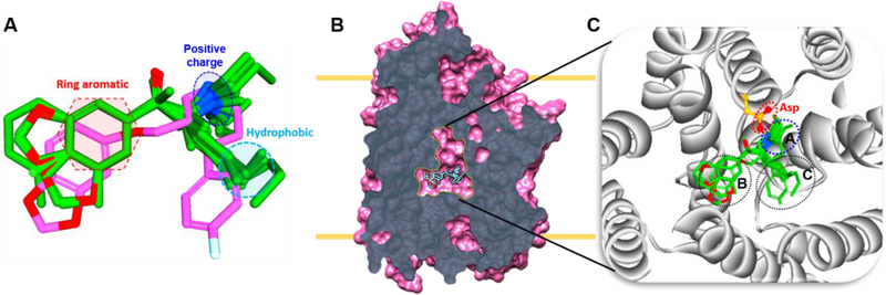
Figure 4.
Alignment of all ligands in the binding pocket. A. Ligand alignment based on the paroxetine binding pose derived from hSERT crystal structure (PDB: 5I6X). Key pharmacophore features such as positive charge, aromatic ring and hydrophobic are shown in the dotted lines. B. Slab view of hSERT and the central site showing aligned ligands. C. An enlarged view of central site and plausible ligand binding poses, dotted lines indicate subsites A, B, and C.

