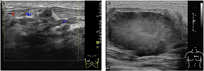
Figure 3.

(a) Ultrasound shows the structure of the mammary gland disorder, skin thickening subcutaneous tissue space edema, and low echo area, suggesting breast cancer (Grade III invasive ductal carcinoma). (b) Ultrasound shows a 5.0 × 2.9 × 4.8 cm hypoechoic, undersmooth, irregular, and lobulated mass, which indicates lobular neoplasms (Grade II invasive ductal carcinoma).
