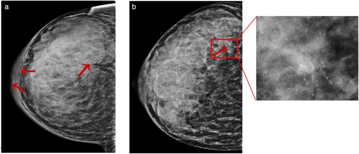
Figure 4.

(a) Mammography shows isodense masses of irregular shape but mostly smooth margins and crater nipples, as well as pachyderma around the mammary areola, which suggests a malignant tumor (Grade III invasive ductal carcinoma). (b). Conventional mammography image displays the high‐density breast with no obvious malignant signs. Architectural distortion and irregular microcalcifications are only shown in magnified mammograms (Grade II invasive ductal carcinoma).
