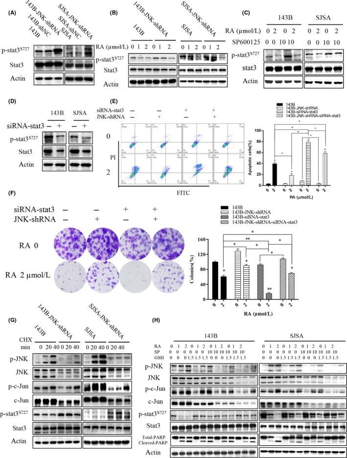
Figure 6.

ROS/JNK/STAT3 pathway mediated Raddeanin A (RA)‐induced apoptosis in human osteosarcoma cells. A‐C, Expression of phospho‐STAT3S727 and STAT3 was analyzed by western blotting. A, Cells treated with shRNA targeting JNK in 143B and SJSA cells. B, Cells treated with RA after JNK knockdown compared with control. C, Cells treated with RA in the presence or absence of SP600125. D‐F, Cells treated with siRNA against STAT3. D, Expression of phospho‐STAT3S727 and STAT3 is detected. E, RA‐induced apoptosis detected in the presence or absence of siRNA‐STAT3 and/or JNK knockdown by flow cytometry. Histograms indicate degrees of apoptosis from three separate experiments. F, Clone formation is detected. Histograms indicate the colony‐formation rate from three separate experiments. G, Cells were incubated in 10 mg/mL CHX (cycloheximide) for various times (0, 20, and 40 min). H, Cells were pretreated with or without GSH/SP600125 for 2 h and then incubated in various concentrations of RA for 48 h. Expression of phospho‐JNK, JNK, phospho‐c‐Jun, c‐Jun, phospho‐STAT3S727, STAT3, and total and cleaved PARP was analyzed by western blotting. GSH, glutathione; PARP, poly‐ADP ribose polymerase; ROS, reactive oxygen species; STAT3, signal transducer and activator of transcription 3
