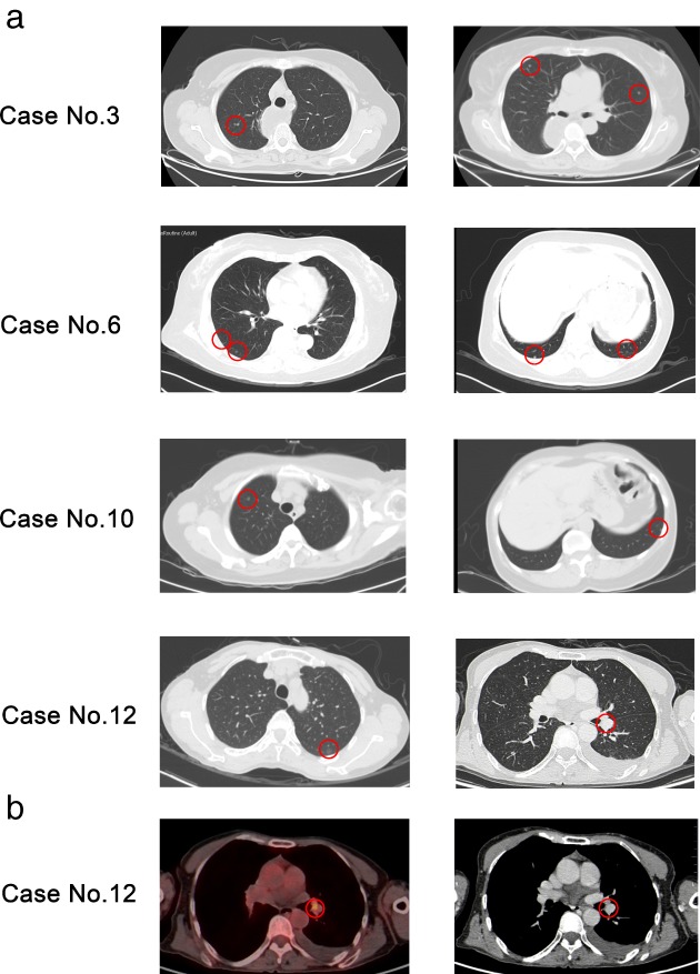
Figure 1.

(a) Thin‐section chest computed tomography scan (1 mm collimation) demonstrated multiple well‐defined nodules of various sizes in four patients. (b) Preoperative 18‐fluoro‐2‐deoxy‐D‐glucose positron emission tomography examination revealed an increased maximum standardized uptake value of 4.8 in one patient. Red circles indicate pathological representation of minute pulmonary meningothelial‐like nodules.
