Figure 3.

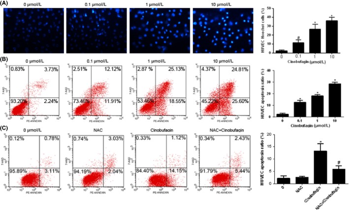
Cinobufagin induces apoptosis in HUVEC. A, Hoechst 33342 staining for HUVEC with cinobufagin treatment (n = 3). Cells were treated with different concentrations of cinobufagin (0, 0.1, 1, and 10 μM) for 24 h. Original magnification, 200×. Marked morphological changes in chromatin morphology such as crenation, condensation, and fragmentation are observed in HUVEC. B, Apoptosis rate of HUVEC cells detected by cytometry (n = 3). Cells treated with different concentrations of cinobufagin (0, 0.1, 1, and 10 μM) for 24 h were detected by flow cytometry. C, Apoptosis rate of HUVEC treated with N‐acetyl‐l‐cysteine (NAC; n = 3). l‐NAC reduced HUVEC cell apoptosis induced by cinobufagin. Cells treated with or without cinobufagin or NAC (0.1 μM) for 24 h were detected by flow cytometry. # P < .05; *P < .01 compared to 0 μM
