-
A–H
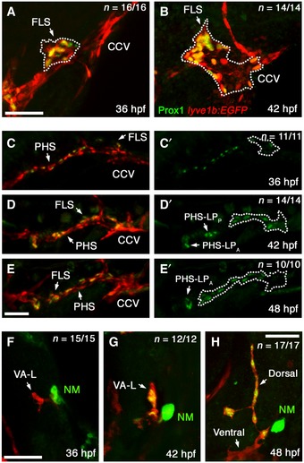
Lateral images of the CCV fixed at 36 hpf (A) and 42 hpf (B), of the PHS at 36 hpf (C), 42 hpf (D) and 48 hpf (E) and of the VA‐L at 36 hpf (F), 42 hpf (G) and 48 hpf (H) in lyve1b:EGFP fish that have been fluorescently immunostained with anti‐PROX1 (green) and anti‐GFP (red). PHS Prox1 staining shown alone for all timepoints (C’–E’) with the FLS demarcated (dotted line) from the adjacent vessels, and the positions of the PHS‐LPP and PHS‐LPA indicated. Note the Prox1‐positive (green), GFP‐negative structure near to the VA‐L is a prox1‐expressing neuromast (NM).
Data information: CCV, common cardinal vein; FLS, facial lymphatic sprout; hpf, hours post‐fertilisation; NM, neuromast; PHS, primary head sinus; PHS‐LP
A, anterior primary head sinus lymphatic progenitor domain; PHS‐LP
P, posterior primary head sinus lymphatic progenitor domain; VA‐L, ventral aorta lymphangioblast. Scale bar = 50 μm.

