-
A–N’
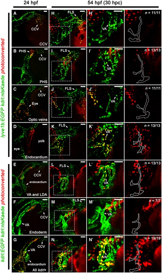
Lateral (A–G) and ventrolateral (H–N) images of the facial vessels in kdrl:nlsKaede;lyve1b:EGFP (A–D, H–K) or kdrl:nlsKaede;kdrl:EGFP (E–G, L–N) embryos. The CCV (A), PHS (B), optic veins (C), endocardium (D), VA, LDA (E), pharyngeal endoderm (F) and all kdrl‐positive tissues in the head (G) were photoconverted red at 24 hpf, while unconverted Kaede, and kdrl:EGFP or lyve1b:EGFP for clarity, is shown in green. All photoconverted embryos were traced to 54 hpf (H–N). Panels (H’–N’) are higher magnification images of the dotted boxes in (H–N) and are accompanied by a red channel only (traced photoconverted cells) image with the VA‐L (dotted line) demarcated from the adjacent VA. No photoconverted kaede‐positive cells seen in the VA‐L in all cases.
Data information: CCV, common cardinal vein; FLS, facial lymphatic sprout; hpf, hours post‐fertilisation; hpc, hours post‐conversion; PHS, primary head sinus; LDA, lateral dorsal aorta; VA, ventral aorta; VA‐L, ventral aorta lymphangioblast. Scale bar = 50 μm.

