
Figure 3.
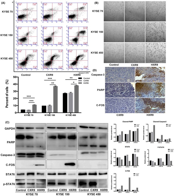
HXR9 induced esophageal squamous cell carcinoma cell apoptosis. A, Effect of HXR9 or CXR9 on cell apoptosis was assessed by flow cytometry using Annexin V/PE after treatment for 2 h. The apoptosis rate for HXR9‐treated cells was significantly higher than that for CXR9‐treated cells. B, Light micrographs of KYSE70, KYSE150 and KYSE450 cells treated with 60 μmol/L CXR9 or HXR9 for 2 h. A great deal of cell shrinkage and pieces of cell debris was observed in HXR9‐treated cells. C, Several apoptosis indicators were analyzed using western blotting. Caspase‐3 and poly ADP ribose polymerase (PARP) activity did not increase significantly over 2 h with HXR9 treatment. However, c‐fos showed significant upregulation in HXR9‐treated KYSE70 and KYSE450 cells. Meanwhile, p‐signal transducer and activator of transcription (p‐STAT)6 was downregulated in HXR9‐treated cells, which was an indicator of anti‐apoptosis. D, In tumors removed from animals, expression of Caspase‐3, PARP and c‐fos showed remarkable upregulation by HXR9 treatment. *P < 0.05, **P < 0.01 and ***P < 0.001